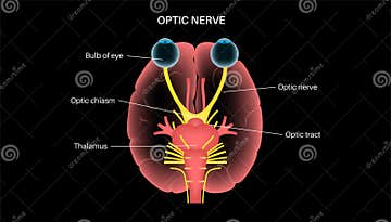
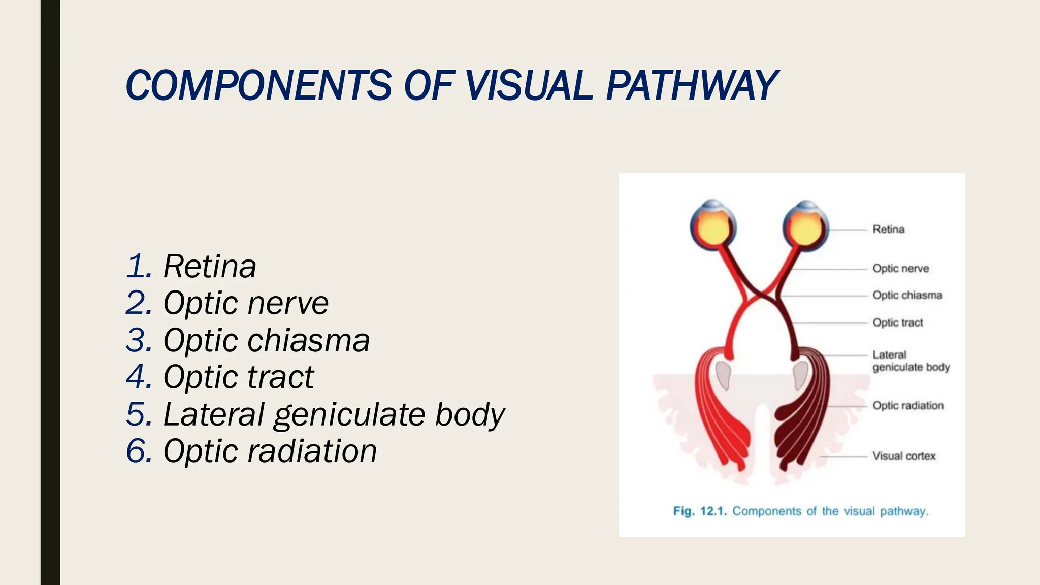
Optical Nerve Scheme Stock Illustration 55036114 Biology Diagrams Download scientific diagram | Schematic of the optic nerve and its different segments. Schematic of the optic nerve and its different segments. from publication: A Narrative Review of Point of Learn about the optic nerve (CN II), the second cranial nerve that transmits visual information from the eye to the brain. See the anatomical course, the optic chiasm, the optic tract and the optic radiation, and their clinical relevance.
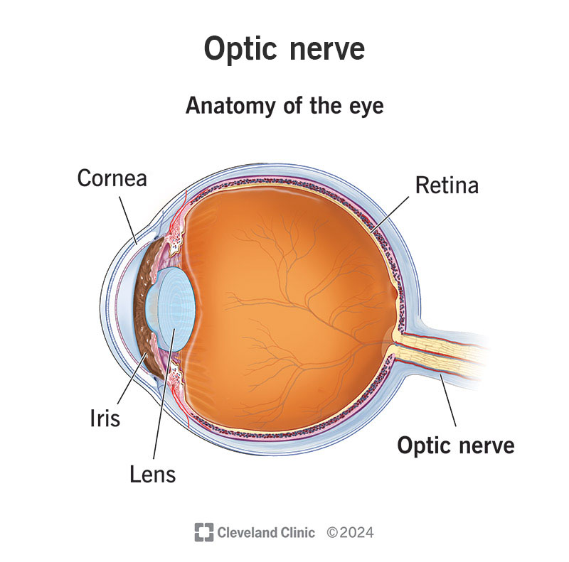
In the center of the retina is the optic nerve, a circular to oval white area measuring about 2 x 1.5 mm across. A simplistic wiring diagram of the retina emphasizes only the sensory photoreceptors and the ganglion cells with a few interneurons connecting the two cell types such as seen in Figure 2. Fig. 2. Simple organization of the retina. Optic nerve anatomy. The optic nerve is located at the very back of the eye, attached to the retina. Because of its function, the optic nerve is considered part of the nervous system, even though it's located in the eye. The Optic Nerve (cranial nerve II) Nerve (ganglionic) cells as well as millions of nerve fibers make up the optic nerve.
Understanding the Optic Nerve: Connecting the Eye to the Brain Biology Diagrams
Learn about the optic nerve, a paired cranial nerve that transmits visual information from the retina to the brain. See the anatomy, function and clinical significance of the optic nerve, as well as its relation to the visual cortex and the eye's blind spot.
Learn about the optic nerve, the connection between your eyes and your brain that lets you see. Find out how it works, where it is, and what can affect it.

Optic nerve Biology Diagrams
The optic nerve has been studied heavily because it is a direct extension of the brain. Anatomy . The optic nerve is mainly made up of the axons (nerve fibers) of the retinal ganglion cells from the retina. The optic disc or nerve head is the point where the axons from the retinal ganglion cells leave the eye. The optic nerve also plays a role in controlling the size of the pupil, the small opening in the center of the iris that regulates the amount of light entering the eye. When the pupil constricts, or becomes smaller, it helps to reduce the amount of light entering the eye, which can be beneficial in bright light conditions. [2] Conversely, when the pupil dilates, or becomes larger, it allows