Regulation of Cell Division PowerPoint Presentation Biology Diagrams The DNA damage checkpoint pathways consist of lesion sensors, adaptors/mediators that assemble signaling complexes at DNA lesions, protein kinases, and their substrate effectors (Table 1). The proteins identified as components of the DNA damage checkpoint pathways are part of the surveillance network that also regulates the orderly progression through an unperturbed cell cycle. Although the G1
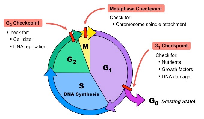
These checkpoints occur near the end of G 1, at the G 2 /M transition, and during metaphase. Figure 10.3B. 1 10.3 B. 1: Internal Checkpoints During the Cell Cycle: The cell cycle is controlled at three checkpoints. The integrity of the DNA is assessed at the G1 checkpoint. Proper chromosome duplication is assessed at the G2 checkpoint.
G1 Phase Cell Cycle Checkpoint Biology Diagrams
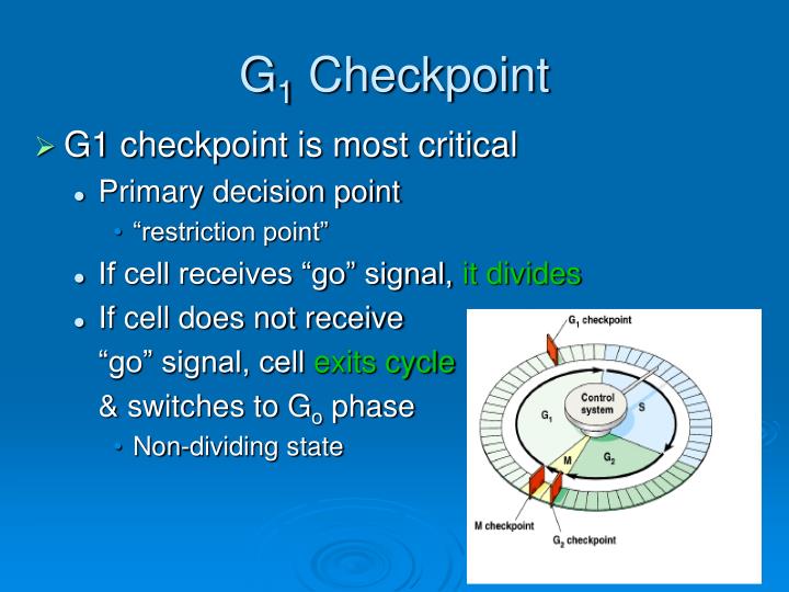
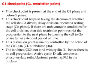
The G1 checkpoint, also known as the restriction point in mammalian cells and the start point in yeast, is the point at which the cell becomes committed to entering the cell cycle. As the cell progresses through G1, depending on internal and external conditions, it can either delay G1, enter a quiescent state known as G0, or proceed past the restriction point. [5] DNA damage is the main

Specializing cells for specific functions. Regulation of the Cell Cycle Checkpoints tightly regulate the cell cycle to prevent errors. These checkpoints include: G1 Checkpoint: This checkpoint ensures that the cell has adequate energy resources and that the surrounding environment is favorable for DNA replication. Cell cycle checkpoints are surveillance mechanisms that monitor the order, integrity, and fidelity of the major events of the cell cycle. These include growth to the appropriate cell size, the replication and integrity of the chromosomes, and their
10.3B: Regulation of the Cell Cycle at Internal Checkpoints Biology Diagrams
Regulation of G1 Cell Cycle Progression Distinguishing the Restriction Point from a Nutrient-Sensing Cell Growth Checkpoint (s)
